Tests and Diagnosis of Pericarditis
Pericarditis is a very "tricky" disease because of both its non-specific, in a layman's opinion, symptoms and its ability to mask as some other disease. If there are any suspicions of pericarditis, it’s necessary to consult a cardiologist. An essential condition for the effective and correct treatment of pericarditis is a proper diagnosis. It is very important to undergo all required tests as soon as possible because pericarditis may be dangerous for the life of a patient.
Diagnostic tests for pericarditis include:
- Taking medical history from a patient, medical examination (auscultation and percussion of the heart).
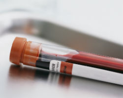
 Blood tests (clinical, biochemical, immunological) to check if there are any signs of inflammation in the blood. For this purpose they evaluate the following blood parameters:
- total protein and its fractions;
- sialic acids;
- fibrinogen;
- seromucoids;
- urea (carbamide);
- creatine kinase.
- Echocardiography (cardiac ultrasound) – it is the main method of pericarditis diagnosis allowing detection of even small amounts of liquid exudate (about 15 ml) in the pericardial cavity, changes in the movements of the heart, adhesions, thickening of pericardial layers.
- Electrocardiogram (ECG) – in the case of pericarditis, the overall electrical activity of the heart is reduced and abrupt decrease in voltage is registered.
- Phonocardiogram (PCG) or recording of heart murmurs – PCG helps register the murmurs that are not associated with normal cardiac cycle and those accompanied with various fluctuations.
- Chest X-ray. Chest X-ray image can show increase in the size as well as changes in the normal configuration of the heart. If adhesions grow in the pericardium, chest X-ray helps detect the so-called "still heart" (it is fixed in one place by these adhesions). If calcium salts are being deposited on the coat of the heart, they form an "armored heart" that is clearly shown on the X-ray image as calcareous deposits on the pericardium.
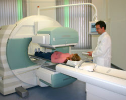
 Computed tomography (CT) – this method shows thickening and calcification (calcium salts deposits) of the pericardium.
- In some cases diagnostic puncture or biopsy of the pericardium are indicated. During these procedures a needle is introduced into the pericardial cavity and the fluid, which is accumulated there, is taken for the further analysis to understand its nature (pus, cancerous cells, fungi, tuberculosis, etc.). It helps make accurate diagnosis and choose treatment tactics.
- MRI of the heart. This procedure is the most sensitive and effective for the detection of pericardium layers thickening.
Next chapter: Treatment and prevention of pericarditis
Featured Articles
A stroke can cause problems with communication if there is damage to the parts of the brain responsi ...
People with irregular bowel movements often complain about elevated blood pressure that doesn’ ...
A mini-stroke is a temporary disruption in blood flow to the brain, spinal cord or retina that doesn ...
It is well known that rheumatoid arthritis is an auto-immune disease that primarily affects joints. ...


