Diagnosis of Heart Defects
In the majority of cases, congenital heart disease can be diagnosed while the woman is still pregnant. After the child is born, the diagnosis is usually confirmed.
Diagnosis During Pregnancy
Fetal echocardiography is the main method used to diagnose congenital heart disease in unborn babies. This method uses an ultrasound scanner to visualize the unborn baby’s heart. Fetal echocardiography should be performed as a part of routine antenatal examination between the 18th and 20th week of pregnancy.
This method, however, can sometimes fail to detect a heart defect, especially if it’s a mild one.
Diagnosis after the birth
If after birth the baby has symptoms which are likely to be caused by congenital heart disease, then this disorder can be identified right away. The most noticeable symptom which can raise a suspicion that the baby might have a congenital heart disease is cyanosis (blue shade of the skin).
Otherwise, the heart defect can remain unnoticed for months and even years.
Further Testing
Additional tests used to diagnose congenital heart disease include:
- Echocardiography – an ultrasound scan that creates in image by using high frequency sound waves. It can be used to visualize all the structures located within the heart and detect any malformations that were missed during the fetal echocardiography.
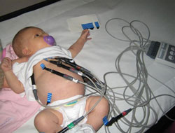
- Electrocardiogram – is a test which detects electrical activity of the heart. The electrodes are placed on the skin at specific points and connected to the computer. The computer then analyzes the signal created by the heart.
- Chest X-ray – can detect whether the heart is larger than normal and whether there is too much blood/liquid within the lungs, which could be signs of heart disease.
- Pulse oximetry – this method can measure the concentration of oxygen within the blood. During this method, a special sensor is placed on the fingertip, toe, or ear. This sensor emits light and detects how this light is absorbed by the tissues. Different amounts of light will be absorbed depending on the concentration of oxygen in the blood.
- Cardiac catheterization – is an invasive, yet informative, method of obtaining more information on the blood flow within the heart. To perform this procedure, a catheter (a narrow tube) is inserted into a blood vessel located in the groin or arm. The catheter is then guided using MRI scanner until it reaches the heart. The end of this catheter can measure blood pressure which can be used to determine the blood pressure within the different parts of the heart. An X-ray positive dye can also be injected through the catheter into the heart to provide a clear picture of the heart chambers.
Next Chapter: Treatment of Congenital Heart defects


